
The magnus is also different due to dual innervation and different blood supply.

The adductor magnus differs as it is considered the most powerful adductor of the thigh the superior horizontal fibers flex the thigh while the vertical fibers extend the thigh.

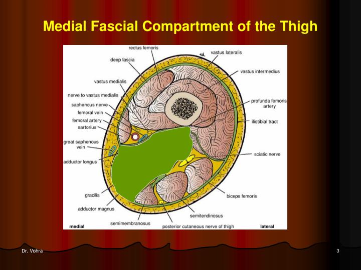
The unique feature of the adductor brevis is that the innervation can be from either the anterior or posterior division of the obturator nerve. The longus is unique because it is innervated by the anterior division of the obturator nerve (L2, 元, L4). The longus and brevis rotate the hip joint laterally and share the blood supply of the obturator and medial circumflex femoral arteries. The longus, brevis, and magnus all adduct and flex the thigh. The adductors of the thigh are the adductor longus, adductor brevis, adductor magnus, and gracillis. Medial Compartment of the Thigh (adductors) All these muscles insert via the common quadriceps tendon into the base and sides of the patella, thus exerting their extension action on the knee joint. The vastus medialis arises from the intertrochanteric line and the linea aspera. The vastus intermedius arises from the anterolateral part of the upper femur. The vastus lateralis arises from the intertrochanteric line, the greater trochanter, and the linea aspera. The rectus femoris has two heads - the reflected head and the straight head - which arise from just above the acetabulum and from the anterior inferior iliac spine, respectively.

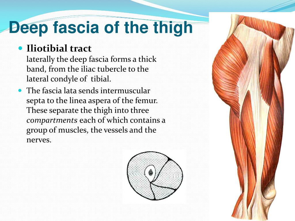
All four function to extend the knee, but the rectus femoris is also an accessory flexor of the hip. The four muscles in the extensor group are the vastus lateralis, vastus intermedius, vastus medialis, and rectus femoris. All muscles in this group are innervated by the femoral nerve (L2-L4) and supplied by the lateral femoral circumflex artery (except the vastus medialis, which receives its blood supply from the femoral artery, profunda femoris artery, and superior medial genicular branch of the popliteal artery. The overall function of the quadriceps group is knee extension via their distal attachments at the patellar tendon. Its blood supply is from the medial circumflex femoral branch of the femoral and obturator artery. The pectineus is also unique as it adducts the thigh and flexes the hip it is usually innervated by the femoral nerve but is sometimes innervated by the obturator nerve. The sartorius is unique in that it flexes and laterally rotates the hip joint and flexes the knee, is innervated by the femoral nerve (L2 through L4), and receives blood supply from the muscular branches of the femoral artery. Innervation to the iliacus is by the muscular branch of the femoral nerve (L1 through 元), while the direct fibers of L1 innervate the psoas through 元 lumbar plexus. The iliacus and the psoas are both flexors of the thigh in relation to the torso and receive blood supply from the lumbar branch of the iliopsoas of the internal iliac artery. The pectineus is a smaller muscle that arises at the eponymous pectineal line of the pubis and inserts at the upper femoral shaft behind the lesser trochanter. The sartorius muscle is the longest in the body and crosses from the anterior superior iliac spine inferomedially to insert at the supper medial part of the tibia. Psoas major arises from the lower border of the T12 vertebra to the L5 vertebra, and it extends downwards under the inguinal ligament with iliacus to inserts beside it at the lesser trochanter of the femur. The iliacus arises from the iliac fossa within the bony pelvis and inserts at the upper part of the femur. The hip flexors are composed of the iliacus, psoas major, sartorius, and pectineus. This compartment includes two subgroups of muscles: those that are hip flexors and hip abductors, and the knee extensors (called the quadriceps collectively) The muscles of the gluteal region are also important to the function of the lower limb but are considered in a separate article. The thigh muscles are divided into three compartments: the anterior, medial, and posterior thigh muscles, each of which contains multiple muscles which broadly share the same functional action, innervation, and arterial supply. Understanding the main function of each group of muscles and the unique properties of muscles within a group will help identify different disorders based on the physical examination and patient history. The thigh muscles are divided into groups based on location – the anterior, medial, and posterior compartments - each affording the lower limb different functional movements. The thigh region is essential to the proper postural stability and locomotion of humans and is also a key conduit for neurovascular structures that pass from the abdomen to the lower limbs.


 0 kommentar(er)
